 Facebook
 Youtube
 Industrial Endoscope
 MENU
MENU
- Adronic minimally invasive fluorescence imaging system
- Medical Endoscope ICG 5 ALA Camera
Adronic Endoscopic 5ALA fluorescence imaging system, is able to observe the precise location of brain tumors and cancer cells. This achieves accurate intraoperative sampling and shorten the operation time and expenses. After the removable of the brain tumors, Adronic ICG fluorescence imaging system assists to observe blood vessels and lymphatic, making sure that blood vessels is not under the risk of injured. In addition, it provides anatomical structure and vital tissues in silhouette in order to achieve an accurate sampling in surgery.
Avoiding carotid artery to be exposed under the risk of injury and operate for various tumors removal. This assist doctors to effectively protect the patient during the operation, and also shorten the operation time and cost. This technology is also the leading technology in the global, and there is no similar product in the market. (Below photos for reference).
Technical Difficulties
Advanced medical endoscopy image quality enhancement technology provides better medical intraoperative accuracy which will be one of the leading technology in the future. A recurring problem that doctors often face is the ability to visualize and predict the relative position of important anatomical structures in a relatively narrow working environment.
In the 20th century, most of the medical clinical operations are done by using microscope. During microsurgery, the operating microscope is only capable of providing views on the superficial structure. With the combination of microscopy technique and endoscope usage, endoscope is able to provide inner part of superficial structure and non-invasive observation.
During the 21st century, doctors have begun using an endoscope to directly perform multiple operations. The market demand for medical endoscopy will continue to rise, and it will drive a steady growth of Adronic Medical Endoscopy.
In addition, the current endoscopic image quality enhancement technology in Europe, America, and Japan is not yet fully developed, and it is difficult to use it in ward, consultation, telemedicine, and in minimally invasive endoscopic surgery.
Furthermore, Adronic has developed an advanced medical endoscopy image quality enhancement technology ICG imaging system, 5 ALA imaging system. If Adronic seizes market opportunities, Adronic will become one of the leading companies.
The technology of advanced medical endoscopy equipment, Adronic will break through the monopoly of Europe, America, and Japan and will definitely increase the impression of Taiwan in the international market.
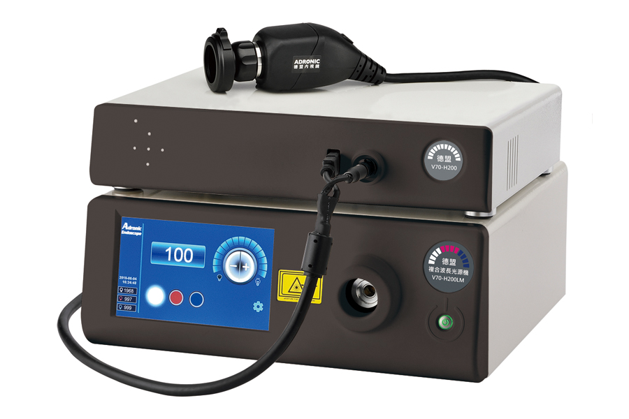
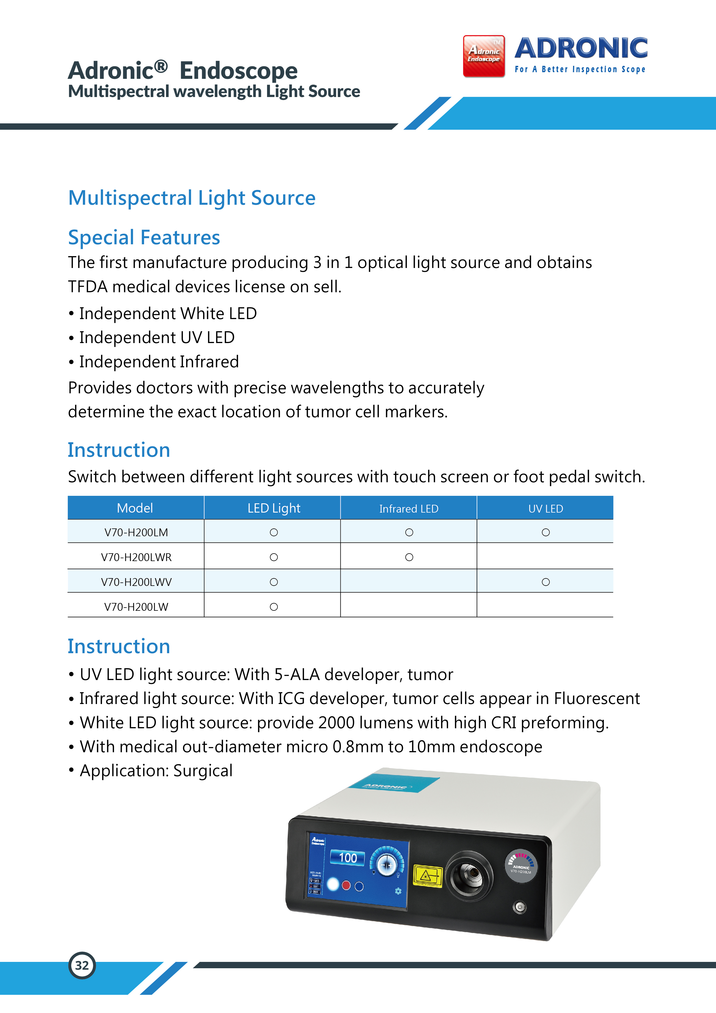
◆ Model:V70-H200LM
◆ The world's only optical independent white light module, blue (UV light) module, and infrared light module.
◆ Matchable with the 5 ALA contrast agents; Through particular wavelengths and fluorescent imaging technology,
the minimally invasive endoscopic photography system can accurately identify the edge of the malignant tumor and the location of the cancer cell.
◆ Matchable with the ICG imaging agent; Through the light source of the same wavelength and fluorescent imaging technology,
the minimally invasive endoscopic photography system can accurately identify the tumor edge, lymphatics, and blood vessels
◆ Multi-stage adjustment of light source intensity
◆ Use the touch panel or pedal to switch the output of different light sources
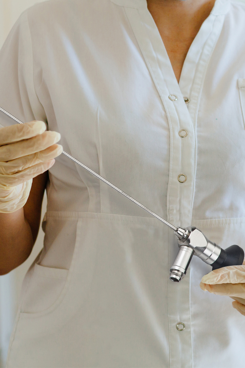
Adronic 3 in 1 Medical Infrared / UV / White Light Source
Multiparametric Endoscopy for the detection of tumor or cancer using wide field multispectral imaging during 5- ALA or ICG
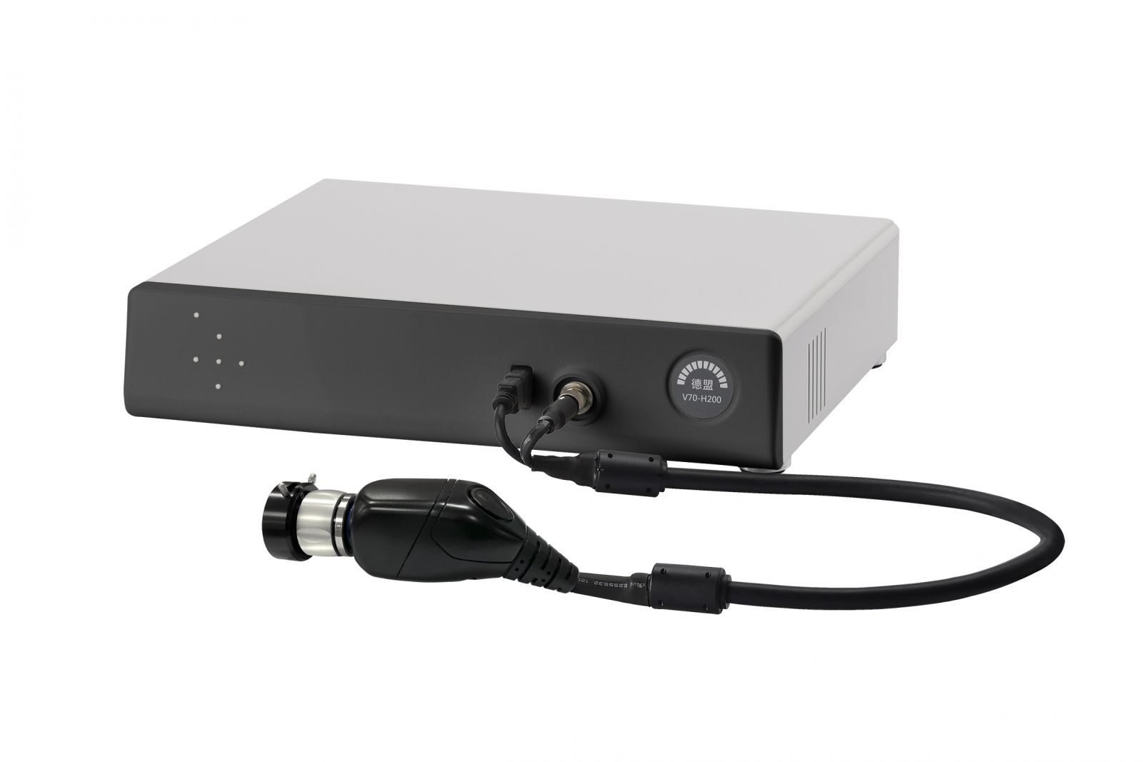
Adronic 4K HD Adronic Station
Adronic 4K HD Adronic Station: Multispectral light scattering endoscopic Fluorescent imaging of Blood vessel and Neurosurgery Tumor.
The unique three independent optical glass spectral design white, blue and IR through foot pedal device which offers a fast and conveniences way that helps the doctor to knows where exactly the tumor cell are located by 5-ALA or ICG contrast medium.
V70-H200LWR When the ICG contrast medium uses infrared wave (IR) that can therefore causes the tumor cells to fluoresce. Doctor will be able to observe where exactly the tumor cells are at.
V70-H200LWV When the 5-ALA contrast medium inject the veins, it can absorb the blue wave, that then causes the tumor show in red from the monitor display. Doctor will be able to observe where exactly the tumor cells are at.
V70-H200LW Ultra-powerful white light source, The image displayed by the endoscope with this light source is excellent. Suitable for Neurological Surgery.
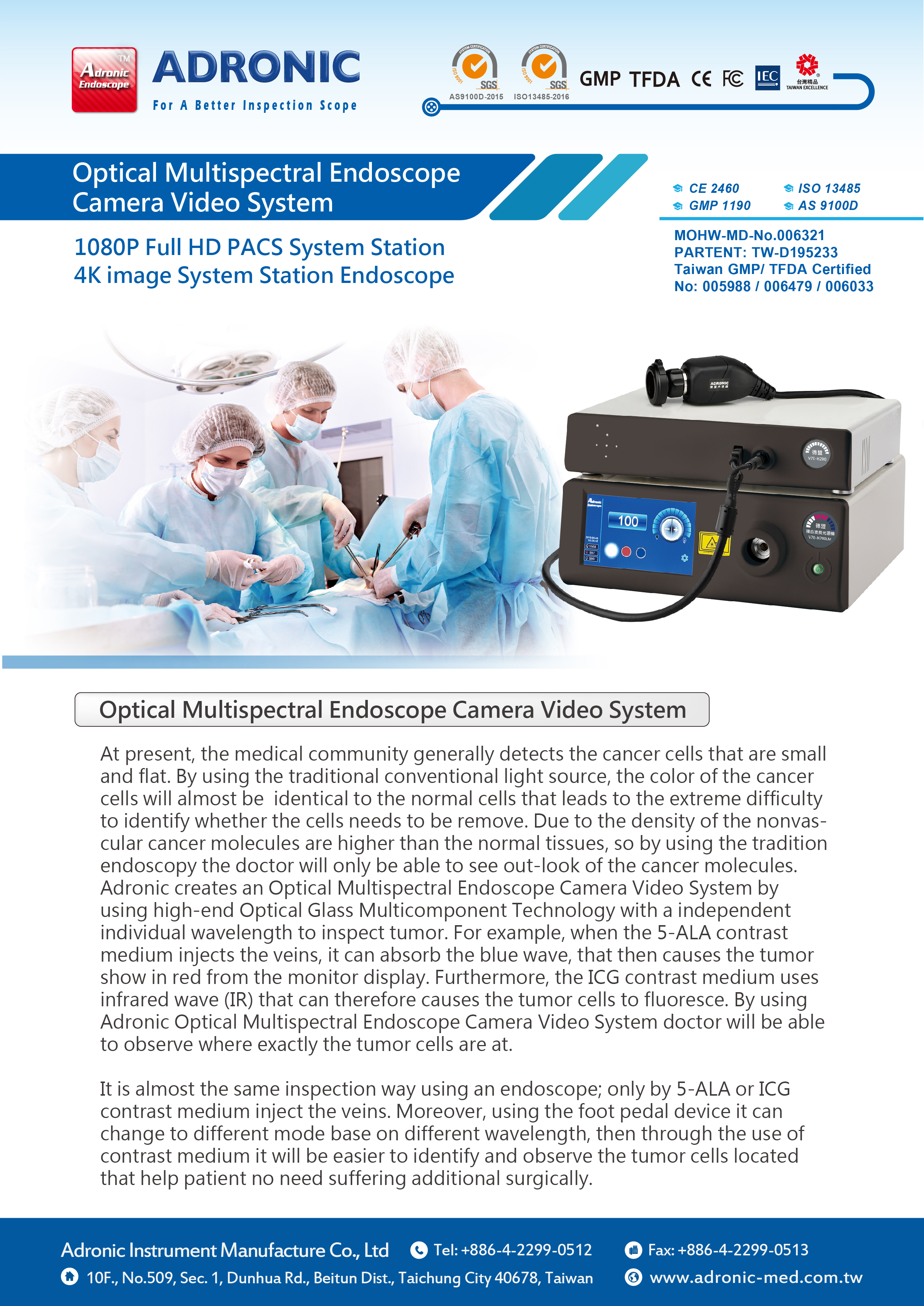
At present, the medical community generally detect the cancer cells that are small and flat. By using the traditional conventional light source, the color of the cancer cells will almost be identical to the normal cells that leads to the extreme difficulty to identify whether the cells needs to be remove. Due to the density of the neovascular cancer molecules are higher than the normal tissues, so by using the tradition endoscopy the doctor will only be able to see out-look of the cancer molecules. Adronic creates an Optical Multispectral Endoscope Camera Video System by using high-end Optical Glass Multicomponent Technology with a independent individual wavelength to inspect tumor. For example, when the 5-ALA contrast medium inject the veins, it can absorb the blue wave, that then causes the tumor show in red from the monitor display. Furthermore, the ICG contrast medium uses infrared wave (IR) that can therefore causes the tumor cells to fluoresce. By using Adronic Optical Multispectral Endoscope Camera Video System doctor will be able to obesere where exactly the tumor cells are at.
The inspection of the Optical Multispectral Endoscope Camera Video System are identical
with the traditional endoscopy, however, by using the Adronic Optical Multispectral Endoscope Camera Video System it provided access to identified the tissue that cannot be easily see. By using the foot pedal device it can change to different mode base on different wavelength, then through the use of contrast medium it will be easier to identify and obser the tumor cells. It can effectively decrease the risks of damaging the normal tissues and the veins inside human body. In addition, it provide the best situation in resecting the sick cells in the save zone that helps the surgeons to make the right move in the operation.
Adronic Optical Multispectral Endoscope Camera Video System includes 4K rigid endoscope, HD endoscope camera with CCU, compounded wavelength optical glass light source and a 4K HD PACS video image system station. 4K rigid endoscope provide crystal image that can identify the difference tumors in the veins that helps the doctor to identify the exact location of the sick cells. 4K HD PACS image capture station helps doctor transfer all images data to main server for necessary records. The unique three independent optical glass spectral design white, blue and IR through foot pedal device which offers a fast and conveniences way that helps the doctor to knows where exactly the tumor cell are located.
Feature
◆ 0.8mm Medical flexible Fiber Scope
◆ Ultra HD 4K image out put
◆ Full HD camera 1080P/60fps resolution
◆ 100% design and made in Taiwan
◆ Unique three independent optical glass spectral white,
blue and IR individual wavelength design.
◆ 100% after sales/repairing services
◆ 7 inch touch panel
◆ Foot pedal allow user to switch White or RED or UV light output.
◆ Advance touch panel allow user to switch White or RED or UV light output
◆ Allow to identify tumor located by 5-ALA or ICG contrast medium inject the veins,
◆ Fast and easy to adjust different light wavelength as well as brightness output level.
Avoiding carotid artery to be exposed under the risk of injury and operate for various tumors removal. This assist doctors to effectively protect the patient during the operation, and also shorten the operation time and cost. This technology is also the leading technology in the global, and there is no similar product in the market. (Below photos for reference).
Technical Difficulties
- At present, the image quality enhancement technology in Europe, America, and Japan uses ICG cameras for microsurgery occupied too much space and cannot be used in ward, consultation and telemedicine.
- At present, 5ALA imaging system is not yet applied in minimally invasive endoscopy.
- Existing endoscopes are mostly wide-angle, and the edges of the shooting images are also distorted. For example, CT and MRI need a lot of preparation, such as: locating the exact location of where the patient lays, making sure the patient and the CT bed, the CT camera, and the camera head is fixed. Fixed elements and apply contrast medium to enhance the accuracy of locating CT/MRI. A wide range brands of endoscopes are designed very differently, and it can greatly affect the accuracy of the imaging. For instance, the image curved surface is too large, and structure of endoscopes will directly affect the accuracy and the quality of the image.
- Adronic 4K rigid endoscope: The image distortion ratio is less than 5%. (Obtaining MOHW Class 2 certificate, CE 2460 Class 2 certificate).
- Adronic endoscope - ICG / 5ALA imaging technology
- Adronic multispectral wavelength light source provide White LED / Infrared LED / UV LED, with the combination of White/ UV LED imaging system and White/ Infrared LED imaging system. Functional endoscopy imaging system provides doctors to navigate through the operational location by using White light source in surgery.
- When the light source is switch to UV light, the tumor and cancer cell will be visible on the monitor if the patient is injected with 5 ALA contrast medium. This assists doctors to operate a inner brain surgery for tumor removal.
- Endoscope imaging system technology includes:
- 4K image resolution, hardware, and software that can receive white light/UV light spectrum imaging.
- Hardware, and software that can be switch between white light and UV light. (MOHW Class 2 certificate)
- White light, Infrared light, UV light, with one output technique (Three-in-one Light Source)
- Light source cable support high temperature tolerance with high technique of light transmission
- The endoscope distortion ration is under 5%
- ICG imaging system contains white and infrared light
- 5 ALA imaging system contains white and UV light technology
- Light source cable heat-resistant temperature up to 250 degrees, light transmission is higher than 50% technique
- Adronic multispectral wavelength medical imaging system includes:
- 4k high-resolution endoscope
- HD endoscope camera
- Multispectral wavelength light source
- 4K HD PACS system image storage working station
- The endoscope apply 4K high-quality with Full HD 1080P/60fps image resolution. This assists doctors to identify the tumor inside the vein, and identifying where exactly the tumor is located.
- 4K HD PACS image storing station helps the doctor in storing information into the main server, and to access the record
- Multispectral wavelength light source design a unique optical fiber with White, Infrared and UV light that can switch between lighting output via foot pedal or touch panel. The intensity of the light source can be adjusted easily and can provide images that can best assist doctors to precisely identify the location of the tumor cell.
- Launch a wavelength of 405/780nm light source to the object
- Inject the 5 ALA/ICG contrast medium into the patient
- 5 ALA(ICG) imaging system utilize fluorescence reaction technique through 5ALA camera (ICG camera),with RGB camera system to calculate tumor , cancer cell appearing in red object ( ICG: blood vessel, lymphatic appears in fluorescence color).
Advanced medical endoscopy image quality enhancement technology provides better medical intraoperative accuracy which will be one of the leading technology in the future. A recurring problem that doctors often face is the ability to visualize and predict the relative position of important anatomical structures in a relatively narrow working environment.
In the 20th century, most of the medical clinical operations are done by using microscope. During microsurgery, the operating microscope is only capable of providing views on the superficial structure. With the combination of microscopy technique and endoscope usage, endoscope is able to provide inner part of superficial structure and non-invasive observation.
During the 21st century, doctors have begun using an endoscope to directly perform multiple operations. The market demand for medical endoscopy will continue to rise, and it will drive a steady growth of Adronic Medical Endoscopy.
In addition, the current endoscopic image quality enhancement technology in Europe, America, and Japan is not yet fully developed, and it is difficult to use it in ward, consultation, telemedicine, and in minimally invasive endoscopic surgery.
Furthermore, Adronic has developed an advanced medical endoscopy image quality enhancement technology ICG imaging system, 5 ALA imaging system. If Adronic seizes market opportunities, Adronic will become one of the leading companies.
The technology of advanced medical endoscopy equipment, Adronic will break through the monopoly of Europe, America, and Japan and will definitely increase the impression of Taiwan in the international market.
◆ Model:V70-H200LM
◆ The world's only optical independent white light module, blue (UV light) module, and infrared light module.
◆ Matchable with the 5 ALA contrast agents; Through particular wavelengths and fluorescent imaging technology,
the minimally invasive endoscopic photography system can accurately identify the edge of the malignant tumor and the location of the cancer cell.
◆ Matchable with the ICG imaging agent; Through the light source of the same wavelength and fluorescent imaging technology,
the minimally invasive endoscopic photography system can accurately identify the tumor edge, lymphatics, and blood vessels
◆ Multi-stage adjustment of light source intensity
◆ Use the touch panel or pedal to switch the output of different light sources
Adronic 3 in 1 Medical Infrared / UV / White Light Source
Multiparametric Endoscopy for the detection of tumor or cancer using wide field multispectral imaging during 5- ALA or ICG
Adronic 4K HD Adronic Station
Adronic 4K HD Adronic Station: Multispectral light scattering endoscopic Fluorescent imaging of Blood vessel and Neurosurgery Tumor.
The unique three independent optical glass spectral design white, blue and IR through foot pedal device which offers a fast and conveniences way that helps the doctor to knows where exactly the tumor cell are located by 5-ALA or ICG contrast medium.
V70-H200LWR When the ICG contrast medium uses infrared wave (IR) that can therefore causes the tumor cells to fluoresce. Doctor will be able to observe where exactly the tumor cells are at.
V70-H200LWV When the 5-ALA contrast medium inject the veins, it can absorb the blue wave, that then causes the tumor show in red from the monitor display. Doctor will be able to observe where exactly the tumor cells are at.
V70-H200LW Ultra-powerful white light source, The image displayed by the endoscope with this light source is excellent. Suitable for Neurological Surgery.
| Model | White light | RED light | UV light |
|---|---|---|---|
| V70-H200LM | ○ | ○ | ○ |
| V70-H200LWR | ○ | ○ | |
| V70-H200LWV | ○ | ○ | |
| V70-H200LW | ○ | ||
| V70-H200LR | ○ | ||
| V70-H200LV | ○ |
At present, the medical community generally detect the cancer cells that are small and flat. By using the traditional conventional light source, the color of the cancer cells will almost be identical to the normal cells that leads to the extreme difficulty to identify whether the cells needs to be remove. Due to the density of the neovascular cancer molecules are higher than the normal tissues, so by using the tradition endoscopy the doctor will only be able to see out-look of the cancer molecules. Adronic creates an Optical Multispectral Endoscope Camera Video System by using high-end Optical Glass Multicomponent Technology with a independent individual wavelength to inspect tumor. For example, when the 5-ALA contrast medium inject the veins, it can absorb the blue wave, that then causes the tumor show in red from the monitor display. Furthermore, the ICG contrast medium uses infrared wave (IR) that can therefore causes the tumor cells to fluoresce. By using Adronic Optical Multispectral Endoscope Camera Video System doctor will be able to obesere where exactly the tumor cells are at.
The inspection of the Optical Multispectral Endoscope Camera Video System are identical
with the traditional endoscopy, however, by using the Adronic Optical Multispectral Endoscope Camera Video System it provided access to identified the tissue that cannot be easily see. By using the foot pedal device it can change to different mode base on different wavelength, then through the use of contrast medium it will be easier to identify and obser the tumor cells. It can effectively decrease the risks of damaging the normal tissues and the veins inside human body. In addition, it provide the best situation in resecting the sick cells in the save zone that helps the surgeons to make the right move in the operation.
Adronic Optical Multispectral Endoscope Camera Video System includes 4K rigid endoscope, HD endoscope camera with CCU, compounded wavelength optical glass light source and a 4K HD PACS video image system station. 4K rigid endoscope provide crystal image that can identify the difference tumors in the veins that helps the doctor to identify the exact location of the sick cells. 4K HD PACS image capture station helps doctor transfer all images data to main server for necessary records. The unique three independent optical glass spectral design white, blue and IR through foot pedal device which offers a fast and conveniences way that helps the doctor to knows where exactly the tumor cell are located.
Feature
◆ 0.8mm Medical flexible Fiber Scope
◆ Ultra HD 4K image out put
◆ Full HD camera 1080P/60fps resolution
◆ 100% design and made in Taiwan
◆ Unique three independent optical glass spectral white,
blue and IR individual wavelength design.
◆ 100% after sales/repairing services
◆ 7 inch touch panel
◆ Foot pedal allow user to switch White or RED or UV light output.
◆ Advance touch panel allow user to switch White or RED or UV light output
◆ Allow to identify tumor located by 5-ALA or ICG contrast medium inject the veins,
◆ Fast and easy to adjust different light wavelength as well as brightness output level.
◆ Light Source Model:
V70-H200LM / V70-H200LWR / V70-H200LWV
V70-H200LW / V70-H200LR / V70-H200LV
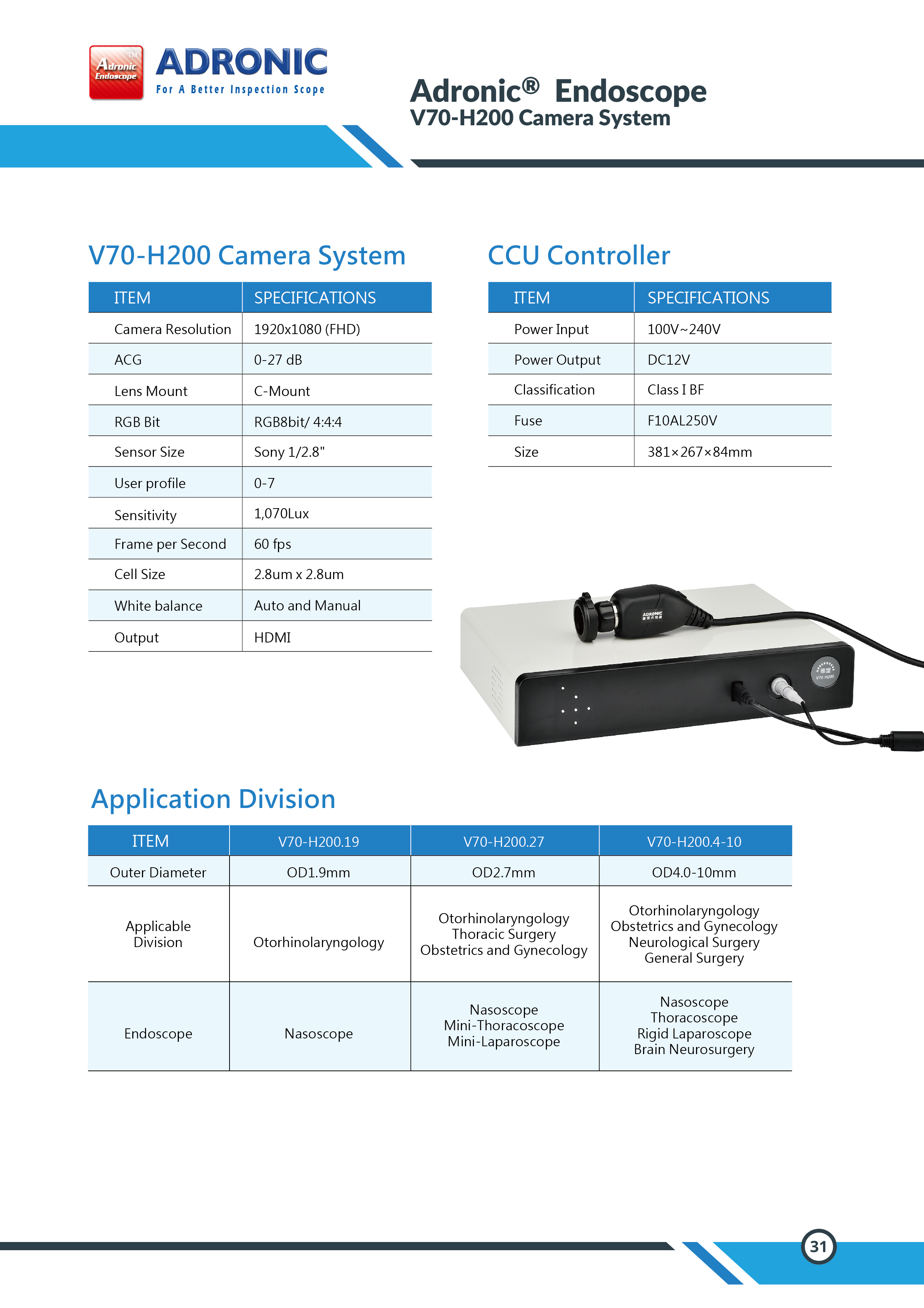
◆ Model: V70-H200
4K/60fps Camera
Full HD2160P/60fps Camera
with Camera Control Unit
V70-H200LM / V70-H200LWR / V70-H200LWV
V70-H200LW / V70-H200LR / V70-H200LV
| V70-H200L Light source |
White light | IR Light | UV light |
|---|---|---|---|
| V70-H200LM | ○ | ○ | ○ |
| V70-H200LWR | ○ | ○ | |
| V70-H200LWV | ○ | ○ | |
| V70-H200LW | ○ | ||
| V70-H200LR | ○ | ||
| V70-H200LV | ○ |
| Light source | V70-H200L |
|---|---|
| Camera Effective Pixels | 1920×1080P |
| Minimum illumination | 0.6lux (AGC ON) F1.2 |
| Lens Mount | C-Mount |
| Bit depths | RGB24bit |
| Sensor Size | Sony CCD 1/2.8〞 |
| User profile | 0-7 |
| Light intensity | >2,000,000LX |
| Life time | >30,000hr |
| LED Color temperature | 6000K |
| Lumen | 2195 Lm |
| Per second photo fram | 60 fps |
| Cell Size(HXV,μm) | 2.8um x 2.8um |
| White balance | Auot and Manual |
| Output | HDMI/VGA |
| Low Carbon | Yes |
◆ Model: V70-H200
4K/60fps Camera
Full HD2160P/60fps Camera
with Camera Control Unit
| Camera | V70-H200 |
|---|---|
| Resolution | 3840+2160 (QFHD) |
| ACG | 0-27 dB |
| Lens Mount | C-Mount |
| RGB Bit | RGB8bit/ 4:4:4 |
| Sensor Size | Sony 1/2.5〞 |
| User profile | 0-7 |
| Sensitivity | 1,070Lux |
| Frame per Second | 60 fps |
| Cell Size | 1.26um x 1.26um |
| White balance | Auot and Manual |
| Output | HDMI |
| Model | Outer Diameter | Applicable Division | Endoscope |
|---|---|---|---|
| V70-H200.08 | OD0.8mm | Gynecology / Urology Division | Cystoscope / Urethoscopy |
| V70-H200.10 | OD1.0mm | Gynecology / Urology Division | Cystoscope / Urethoscopy |
| V70-H200.1.9 | OD1.9mm | Ears, Nose, and Throat(Pediatrics) | Nasoscope |
| V70-H200.2.7 | OD2.7mm | Ears, Nose, and Throat / Thoracic Surgery / Gynecology | Nasoscope / Thoracoscopy / Laparoscopic |
| V70-H200.4-10 | OD4.0-10mm | Ears, Nose, and Throat / Gynecology Neurological Surgery / General Surgery |
Nasoscope(4mm) / Laparoscopic(10mm) / Hard laparoscopic(10mm) / Brain neurosurgery |
.jpg)
